logo-page-magnus

DMSO-FREE CRYOPROTECTANT HARNESSING NATURE AND TECHNOLOGY.

Magnus provides key elements ideal for bone graft formation:

  • An osteoconductive three-dimensional scaffold with cortical and cancellous components.
  • A demineralized bone scaffold with osteoinductive potential.


Does Particle Size Make a Difference?

magnus-img01

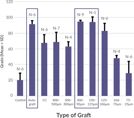
Magnus bone matrix provides a natural, osteoconductive bone scaffold composed of demineralized cortical and cancellous bone. The optimized microparticulate bone scaffold size range of 100-300 μm has been shown to induce simultaneous activity of osteoclasts and osteoblasts, demonstrating rapid healing of bone defects.

100-300 μm optimized particle size for bone regeneration has been shown to support direct ossification, with defect healing rates comparable to autograft.

selector
100-300 μm optimized particle size for bone regeneration has been shown to support direct ossification, with defect healing rates comparable to autograft.

Post-Thaw Cell
Characterization

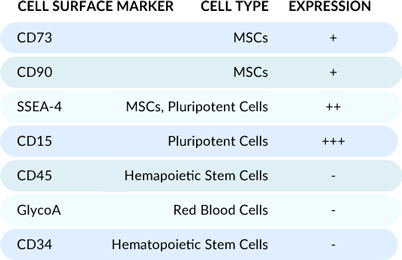
Flow cytometry analysis of the cells post-thaw reveals a unique cell population optimal for osteogenic supplementation, evidenced by high expression of MSC and pluripotent cell markers.

magnus-img02

Cell Preservation:
A Different Approach

magnus-ball-01

DMSO-free cryoprotectant coats the cells to prevent crystalline damage.

magnus-ball-02

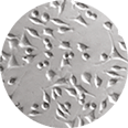
Magnus Representative Sample

magnus-ball-03

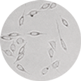
2.5% DMSO Media Sample

Industry standard DMSO penetrates and dehydrates the cell from within to prevent crystal formation. At room temperature, DMSO-based cryoprotectants raise concerns about cytotoxicity and negative effects on cell differentiation.


A Growing Body Of Evidence

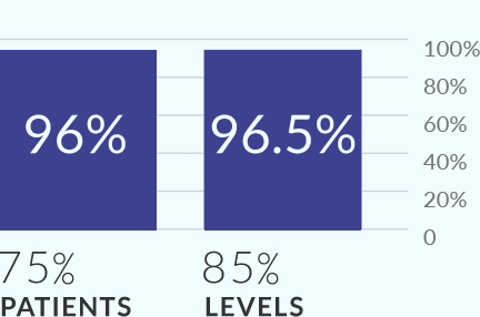
MIS-TLIF study demonstrated 96% fusion at 12 months.
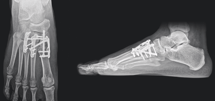
TLIF FUSION with Allograft.  blue-arrow-2020

magnus-img03

It All Adds Up To:

  • A natural scaffold that provides an optimal microenvironment for osteogenesis.
  • A proprietary DMSO-free cryoprotectant that allows for consistent delivery of allograft to the patient.
  • A unique cell population optimal for osteogenic supplementation confirmed through flow cytometry analysis.

Operating Room Ease of Use

  • No rinsing or decanting steps required
  • Minimum of 150,000 cells per cc of allograft5


Midfoot Arthrodesis: Patient with Multiple Comorbidities

12-week post-operative X-rays show complete osseous consolidation of the arthrodesis sites and no pain or swelling with palpation or manipulation of the surgical sites.

magnus-img04


Magnus Allograft Robust, fibrous moldability

Cortical shavings, crushed cancellous chips, and demineralized cortical bone microparticulate scaffold blend with bone gel mixture

Hydrophobic properties make it more resistant to lavage.

available sizes

magnus-img05

References:

  1. Malinin, T.I., et. al., Particulate bone allograft incorporation in regeneration of osseous defects; importance of particle sizes. The Open Orthopeadics Journal, 2007. 1:19-24
  2. Best, Benjamin. P. Cryprotectant Toxicity: Facts, Issues, and Questions. Rejuvenation Research, 2015. Vol. 18, No.5.
  3. Renzi, S., et al., Mesenchymal stromal cell cryopreservation. Biopreservation and Biobanking, 2012. 10(3): p. 276-281.
    Asghar, W., et al., Preserving human cells for regenerative, reproductive, and transfusion medicine. Biotechnology Journal, 2014. 9: p. 895-903.
  4. Data on file at Vivex Biomedical, Inc.
  5. Tally, William C, et al., Transforaminal Lumbar Interbody Fusion with Allograft: 75 Consecutive Cases at 12-Month Follow-Up. International Journal of Spine Surgery, 2018. Vol. 12, No. 1 pp 76-84.