logo-amnio-maxx

Amnio-Maxx is a family of amnion patch allografts that may be used as an anatomical barrier in numerous clinical applications. The natural properties of amniotic tissue help provide mechanical protection to damaged tissue, while the Amnio-Maxx proprietary process retains nutrient-rich growth factors essential for signaling.


About Amnio-Maxx

Growth Factors released from amnio-maxx uc after 24 hours at 37°C

amnio-maxx-img01

selector

Amniotic membrane is a semi-transparent and resilient membrane that lines the upper cavity of the placenta

Amniotic tissue acts as an immune-privileged protective barrier during fetal development1

Amnio-Maxx is applied as an anatomical barrier that helps provide mechanical protection while retaining endogenous growth factors1,2,4

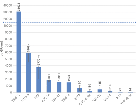
The Amnio-Maxx proprietary process preserves the natural properties of amniotic tissue, maintaining inherent levels of key extracellular matrix molecules, including proteins, carbohydrates, growth factors, and cytokines5

amnio-maxx-img02

Proprietary dual layer technology

  • Dual layer amnion allograft
  • Derived from the amnion layer of the placental membrane
  • Approximately 2X thicker than traditional single layer amnion
  • Available in large sizes for a wide variety of applications

amnio-maxx-img03

Preserving an array of Endogenous growth factors

  • Maximum natural thickness allograft
  • Derived from the umbilical cord
  • Approximately 8X thicker than traditional single layer amnion
  • Offers excellent handling characteristics and the ability to be sutured

Amnio-maxx
orientation diagram

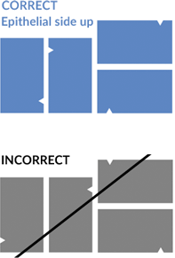
Proper orientation for Amnio-Maxx is achieved when the epithelial layer is facing upwards and the stromal layer is facing downwards, as indicated by:

1. a triangular notch positioned in the upper left hand corner of the allograft (as shown below)

2. An orientation indicator sticker located on the inner pouch facing upwards.

amnio-maxx-img04


Safety and Versatility

  • Amniotic tissue is recovered from healthy mothers who have undergone Cesarean section delivery
  • Amnio-Maxx is processed in accordance with FDA regulations and AATB standards
  • Amniotic tissue has been used for over 100 years with well-documented clinical success3

Amnio-Maxx offers increased versatility across a variety of physician preferences

  • Requires no up-front preparation
  • Hydrates rapidly in the surgical site
  • Ambient temperature storage with a 5-year shelf-life
  • Notch and orientation stickers to designate placement of the epithelial side upwards
  • E-Beam sterilization provides sterility assurance level (SAL) of 10-6

Potential Clinical Applications

  • Spine & Neurosurgery
  • Wound Care
  • Dermatology
  • Oral Surgery
  • Foot & Ankle
  • Burn Care
  • Ophthalmology

References:

  1. Rowlatt, U. (1979). Intrauterine wound healing in a 20-week human fetus. Virchows Arch A Pathol Anat Histol, 381(3), 353–361.
  2. Coolen, N.A. et al. (2010). Comparison between human fetal and adult skin. Archives of Dermatological Research, 302(1), 47–55.
  3. Fairbairn, N.G. et al. (2014). The clinical applications of human amnion in plastic surgery, 67, 662-675.
  4. Niknejad H, Peirovi H, Jorjani M, et al. Properties of the amniotic membrane for potential use in tissue engineering. Eur Cell Mater. 2008;15:88-89.
  5. Delcroix GJ, Namin S, D’Ippolito G, Temple HT, Marshall R. Preserving the natural regenerative potential of amniotic membrane. Vivex Biomedical.
  6. Data on file at Vivex Biologics, Inc.